comprehensive imaging services at ofc
Advanced Imaging for Better Orthopedic Care
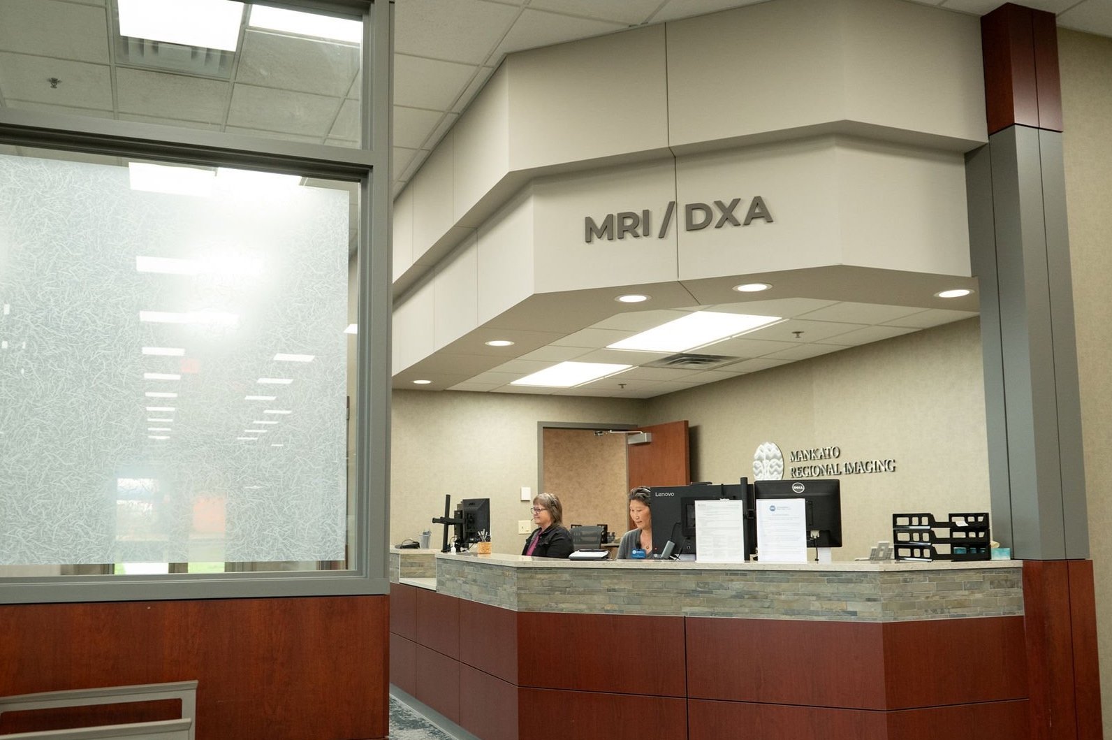
At Orthopaedic & Fracture Clinic, our on-site imaging services—including digital X-ray, MRI, and DXA bone density scans—offer patients convenient, same-day access to advanced diagnostic tools all under one roof. Using the latest technology, our expert team delivers fast, accurate results to support timely diagnosis and treatment. With everything you need in one location, your care is more efficient, streamlined, and personalized from the start.
digital x-ray at ofc
Our state-of-the-art digital X-ray technology provides high-resolution images with faster results. This allows our specialists to quickly and accurately diagnose bone and joint conditions right at the point of care.
advantages of digital x-ray
Faster Images: X-ray results are processed almost instantly, enabling quicker diagnoses and reduced waiting times.
Enhanced Visualization: Digital images can be enhanced, manipulated, and zoomed on a computer screen, providing detailed views of specific areas of interest.
Electronic Storage and Retrieval: Digital images are stored electronically, simplifying patient record management and allowing for easy transfer between healthcare providers.
Remote Access and Consultation: Digital X-rays can be shared electronically, enabling remote consultations with specialists or obtaining second opinions.
Digital Measurement Tools: Advanced measurement and analysis tools in digital X-ray systems help doctors assess angles, distances, and other parameters accurately, aiding in precise treatment planning.
conditions digital x-rays can diagnose
Bone Fractures: Detect fractures with high clarity.
Joint Conditions: Diagnose arthritis and joint abnormalities by analyzing bone spacing and identifying bone spurs.
Tumors: Identify bone tumors and structural abnormalities.
Infections: Detect signs of bone infections, such as osteomyelitis, through changes in bone density and soft tissue swelling.
Orthopedic Conditions: Assess musculoskeletal issues like scoliosis, kyphosis, and other spinal deformities.
Joint Injuries: Evaluate joint injuries such as dislocations or misalignments.
preparing for your x-ray
To ensure a smooth and efficient experience, please bring the following items to your X-ray appointment:
Your doctor’s X-ray order and any related paperwork
Your insurance card(s)
Any previous X-ray files or films of the affected area (if applicable)
Contact Us for More Information
For more information about our Digital X-ray imaging services, call us at 507-386-6600. Our team is here to provide accurate diagnoses and expert care to help you on your journey to better health.
MRI AT OFC
MRI overview
As part of OFC's commitment to delivering exceptional bone, joint, and muscle care, we offer advanced Magnetic Resonance Imaging (MRI) services. MRI technology provides detailed, three-dimensional images of soft tissues, ligaments, and internal structures, enabling our specialists to make precise diagnoses and develop targeted treatment plans. This sophisticated imaging technology helps us deliver the highest standard of orthopedic care while ensuring patient comfort and safety.
What Is MRI?
Magnetic Resonance Imaging (MRI) combines a powerful magnet, an advanced computer system, and radio waves to produce highly detailed images of organs and tissues, helping diagnose a wide range of medical conditions. This non-invasive procedure is accomplished without using radiation or radioactive substances.
MRI Options at OFC
Our facility provides the comfort and convenience of both Large Bore (wide opening) and High Field Open-Sided MRI scanners under one roof, ensuring a positive experience for all patients.
1.2T Hitachi High Field Open-Sided MRI
Our open-sided MRI offers excellent image quality while providing a more spacious environment, making it a great option for patients who are claustrophobic or have a high body mass index.
3T Siemens Large Bore MRI
Ideal for assessing joint injuries and identifying ligament tears, the 3T MRI delivers the most detailed images available. With this cutting-edge technology, our orthopedic experts can develop precise, targeted treatments to improve patient outcomes.
What to Expect During the Examination
You will lie comfortably on the scanning table, and a special coil will be placed near the area being examined. Relax as the scanner and technologist handle the process. You can communicate with the technologist during the exam using a call button and headset/speakers.
During the scan:
You will hear loud knocking noises as the machine captures images.
It is essential to remain still and avoid any movement or coughing.
Headphones or earplugs will be provided. If you choose headphones, you can listen to any Sirius XM satellite radio station.
The average MRI scan takes 30 to 45 minutes.
Getting Your Results
Your MRI images will be interpreted by subspecialty, board-certified radiologists. The reports will be promptly sent to your healthcare provider, who will discuss the results with you in-office or by phone.
Exceptional Care Starts With Extraordinary People
Our technologists have over 65 years of combined MRI imaging experience. Along with our professional, compassionate staff, they are committed to making your visit as relaxed and comfortable as possible.
MRI Accreditation
OFC is accredited by RadSite, a recognition that reflects our commitment to the highest standards of advanced diagnostic imaging.
Patient Safety
Your safety is our highest priority. Since the MRI machine is ALWAYS on, you will be asked to remove all metal objects and complete an MRI safety form before your scan.
Please note: You may not qualify for an MRI if you have any of the following conditions:
Cardiac pacemaker
Brain aneurysm clips
Cochlear, inner, or middle ear implants
History of an eye injury involving a metallic object or foreign body (*If applicable, skull X-rays are required to rule out any remaining metal near the eye.)
Preparation for Your MRI
No special preparation is typically required unless specified by your doctor. You may eat normally and continue taking prescribed medications. Depending on your doctor’s requirements, a contrast agent may be administered intravenously to enhance the visualization of certain structures.
Clothing: Wear loose, comfortable clothing without zippers, buttons, or metal.
Remove all metal objects, including:
Watches, jewelry, eyeglasses
Wallets, purses, credit cards
Cell phones
Hairpins
Dentures
Hearing aids
A secure locker will be provided for your personal belongings.
Important Reminders Before Your Scan
Do not bring any metal objects into the MRI scan room.
Please use the restroom before your scan, as movement is not allowed during the procedure.
DXA AT OFC
DXA (Dual-Energy X-ray Absorptiometry)
At OFC, we offer our patients access to DXA technology as part of our comprehensive range of Imaging services.
Using DXA as a measurement tool within our Bone Health Clinic
DXA is most often used to diagnose osteoporosis, a condition that often affects women after menopause but may also be found in men and rarely in children. Osteoporosis involves a gradual loss of bone, as well as structural changes, causing the bones to become thinner, more fragile and more likely to break.
DXA is effective in tracking the effects of treatment for osteoporosis and other conditions that cause bone loss.
The DXA test can also assess an individual's risk for developing fractures. The risk of fracture is affected by age, body weight, history of prior fracture, family history of osteoporotic fractures and lifestyle issues.
If you think you might have a loss of bone density, a DXA test can be ordered through a referring provider. For more information, please call 507-386-6750.
Using DXA as measurement tool for Health & Wellness
When used for body composition, Orthopaedic & Fracture Clinic’s DXA Scanner can be used by anyone! The process is quick (around 15 minutes) and painless (a scanner hovers over your body while you lay on a table). A DXA scan gives you detailed, precise data about your body fat and muscle composition, and lets you track changes in body composition over time.
Fitness enthusiasts, athletes, and dieters can use body composition scans to get a baseline of where they are now to measure their progress.
Body Composition/Weight Loss
A DXA scan helps you monitor your weight loss progress. A baseline scan gives you your starting point for your body fat, including the amount of unhealthy visceral fat. Subsequent scans allow you to track how much body fat you are losing, and how much muscle you are retaining to keep you moving towards your goals.
Athletes & Fitness
As well as using a DXA scan for basic body fat measurement, athletes often use the scan to pick up muscle asymmetries between their left and right sides. Muscle asymmetries are often caused when our sport causes us to favor one side, or when we use improper form that makes our dominant side do more work.
Health & Wellness
Getting a baseline measurement of fat and muscle let you see where you are today and track changes over time. We often lose muscle and bone mass as we age, making us at greater risk of injury.
Our DXA scan services for Health & Wellness are open to the public without referral. To set up an appointment for your own personal scan or for any questions, please call 507-386-6750.


